13-Week Twin Pregnancy Ultrasound: Chorionicity, Risk Assessment, and First Trimester Milestones
Decoding the Vital Scan: Establishing the Framework for Safe Twin Gestation.
Introduction: The Significance of the 13-Week Twin Scan
The 13-week ultrasound marks a defining moment in any twin pregnancy, serving as the final, crucial opportunity in the first trimester to gather essential data that dictates management for the remaining seven months. At this stage, the embryos officially transition to fetuses, having completed the most vulnerable phase of organogenesis. For expectant parents, this scan offers deep reassurance as the risk of miscarriage drops significantly. For the specialist, the primary, non-negotiable goal is to determine **chorionicity and amnionicity (C/A)**—the structure of the placentas and sacs—which is the single most important factor influencing twin pregnancy risk.
Understanding this ultrasound is about grasping three simultaneous objectives: verifying viability, assessing structural development via the Nuchal Translucency (NT) scan, and, most critically, classifying the twins’ shared environment. This comprehensive guide breaks down the biological shifts and the specific medical information collected at this pivotal early second-trimester appointment.
Fetal Milestones: Growth and Development at 13 Weeks
By 13 weeks, both twins are tiny, active human beings. Organ formation is complete, and the focus shifts entirely to growth, maturation, and functional development.
Size, Measurements, and Emerging Activity
Each fetus is typically the size of a **peach** or a **large plum**. The primary measurement taken is the **Crown-Rump Length (CRL)**, measuring the distance from the top of the head to the bottom of the buttock. This measurement provides the most accurate estimation of gestational age in the first trimester.
- Length: Approximately $2.5 \text{ to } 3.0 \text{ inches}$ ($6 \text{ to } 7.5 \text{ cm}$).
- Weight: Roughly $0.8 \text{ to } 1.0 \text{ ounce}$ ($22 \text{ to } 28 \text{ grams}$).
The twins demonstrate constant, vigorous activity—kicking, curling, swallowing, and even hiccupping—though these movements are still too light for the mother to detect. The ultrasound reveals complex movements, confirming neurological development is on track.
Organ Refinement and Functional Development
Structurally, all systems are in place, but refinement continues:
- Digestive System: The intestines, which began developing outside the abdominal cavity, have now fully retracted into the abdomen.
- Kidneys: The kidneys start producing urine, which is released into the amniotic fluid. The fetus swallows this fluid, which helps train the digestive and urinary systems.
- Vocal Cords: Vocal cords are fully formed, though they will not be used until birth.
- Hands and Fingers: Fingernails are developing, and the twins have the ability to form fists and suck their thumb, behaviors often visible during the scan.
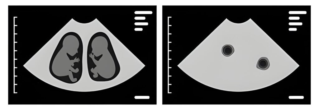
The Primary Goal: Chorionicity and Risk Stratification
The determination of chorionicity—whether the twins share a placenta (chorion) and amniotic sac (amnion)—is the most critical data point gathered at the 13-week scan. This classification dictates the frequency of monitoring and the specific risks associated with the pregnancy.
The Three Vital Classifications (C/A)
The $11\text{ to }14$-week window is optimal for C/A determination, as the dividing membranes are thick and clearly visible. After $14$ weeks, the membranes thin, making accurate diagnosis significantly more challenging.
Twin Classifications and Monitoring Frequency
| Type (C/A) | Placentas / Sacs | Risk Level | Monitoring Frequency (Example) |
|---|---|---|---|
| Di/Di (Dichorionic-Diamniotic) | Two Placentas / Two Sacs | Lowest Risk | Ultrasound every 4 weeks |
| Mo/Di (Monochorionic-Diamniotic) | One Placenta / Two Sacs | High Risk | Ultrasound every 2 weeks |
| Mo/Mo (Monochorionic-Monoamniotic) | One Placenta / One Sac | Highest Risk | Daily/weekly monitoring, likely hospitalization |
The Twin-to-Twin Transfusion Syndrome (TTTS) Risk
The shared placenta in Monochorionic (Mo/Di and Mo/Mo) twins carries the risk of TTTS. This occurs when blood vessels connect on the placenta, causing one twin (the donor) to pump blood to the other (the recipient). The NT scan is the first opportunity to screen for early signs of TTTS, which involves checking the fluid levels in each sac and comparing the sizes of the babies.
The Nuchal Translucency (NT) Scan
Performed specifically between 11 weeks and 13 weeks, 6 days, the NT scan is a crucial component of the first-trimester screening for chromosomal and structural issues. It measures a fluid-filled space at the back of the baby’s neck.
Understanding the NT Measurement
The specialist measures the width of this clear space. While a larger measurement does not definitively diagnose an issue, it correlates with a higher risk for conditions like Down syndrome (Trisomy 21) or congenital heart defects. In a twin pregnancy, this measurement must be taken for each fetus independently, often revealing different risk profiles for each baby.
First Trimester Combined Screening
The NT scan is combined with a maternal blood test that measures two proteins: Pregnancy-Associated Plasma Protein-A (PAPP-A) and free Beta-hCG. Together, these measurements generate a highly sensitive risk factor score for each twin. If the risk is elevated, Non-Invasive Prenatal Testing (NIPT) or diagnostic testing (CVS or amniocentesis) may be recommended.
Specialist Tip: The Lambda Sign
The presence of a **Lambda (or Twin Peak) sign** at 13 weeks strongly indicates a Dichorionic (Di/Di) pregnancy. The absence of this sign, showing only a thin membrane inserting perpendicularly, suggests a Monochorionic (Mo/Di or Mo/Mo) pregnancy. This visual clue guides the specialist's risk assessment.
Maternal and US Socioeconomic Considerations
Carrying twins places significantly higher demands on the mother's body and her environment. At 13 weeks, the mother should be preparing for accelerated changes.
Symptom Changes and Accelerated Weight Gain
Similar to single pregnancies, the severe nausea and fatigue of the first trimester usually peak around now and begin to fade. However, weight gain expectations are higher for twins. General recommendations suggest gaining up to $20 \text{ pounds}$ by 20 weeks for healthy twin pregnancies. This higher caloric requirement is crucial for preventing preterm birth and ensuring adequate fetal growth.
The estimated caloric increase needed by the second trimester for twins is roughly $600 \text{ calories}$ daily above pre-pregnancy maintenance needs, compared to the $340 \text{ calories}$ needed for a singleton pregnancy. This exponential demand drives the need for diligent nutritional planning.
Access to Specialist Care and Financial Impact (US Context)
Diagnosing a Monochorionic twin pregnancy at 13 weeks means the mother requires specialized care from a Maternal-Fetal Medicine (MFM) specialist. Access to these highly specialized doctors and the frequent bi-weekly or weekly ultrasounds they mandate can pose a significant financial burden, even with good insurance coverage in the US healthcare system. Families must proactively investigate their insurance coverage for high-risk obstetric care and MFM consultations immediately after the 13-week scan confirms the twin type.
Interactive Element: Risk-Based Resource Comparison
This tool illustrates the stark difference in monitoring needs based on chorionicity, impacting time, cost, and psychological stress:
Monitoring Comparison (Weeks 16 to 32)
For Di/Di twins, estimated ultrasounds (Weeks 16-32): 5
Conclusion: Proactive Partnership
The 13-week twin ultrasound is more than a viewing; it is the establishment of the entire care plan. By successfully navigating the NT scan and accurately diagnosing chorionicity, you and your medical team gain the framework necessary to manage the specific risks associated with your pregnancy type. Acknowledging the increased demands for monitoring, nutrition, and specialist access allows you to transition into the second trimester with confidence and proactive intent, setting the best possible stage for the safe and healthy growth of both babies.
References and Further Reading
- American College of Obstetricians and Gynecologists (ACOG). (2019). Multifetal Gestation: Twin, Triplet, and Higher-Order Multifetal Pregnancies.
- Society for Maternal-Fetal Medicine (SMFM). (2020). Diagnosis and management of twin-to-twin transfusion syndrome.
- Sperling, D. (2021). High-Risk Pregnancy: Management and Delivery Protocols. Cambridge University Press.


