3D Ultrasound in Pregnancy: Purpose, Safety, and Optimal Timing for Imaging
Differentiating advanced imaging from routine care and managing expectations for elective viewing.
Article Roadmap
2D vs. 3D vs. 4D: Understanding the Technology
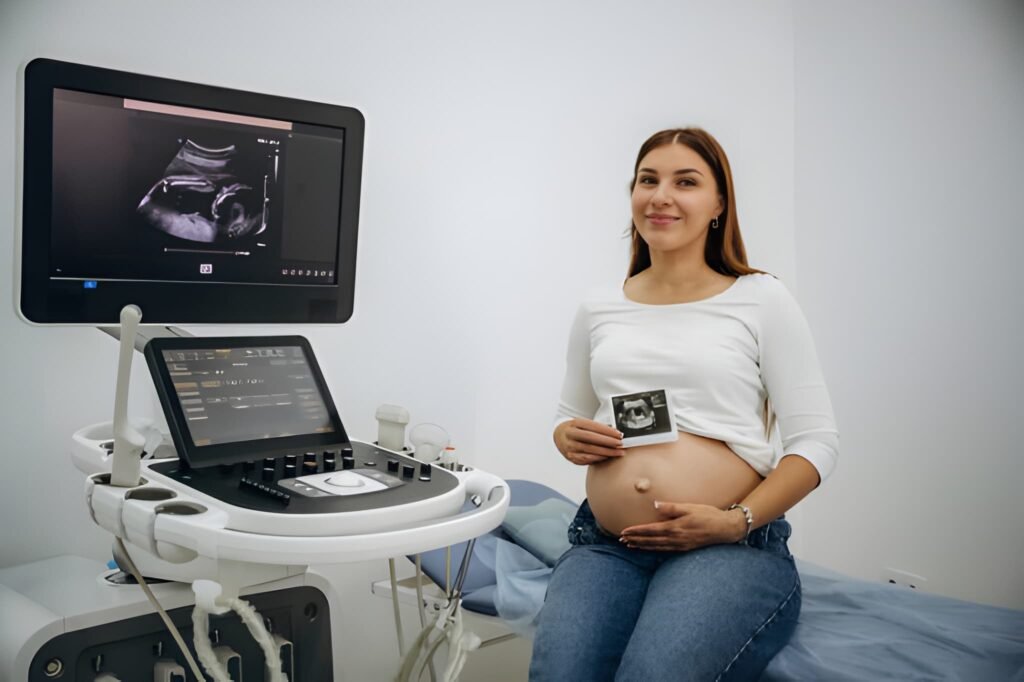
The 3D ultrasound represents an advancement over the traditional scan, providing a depth perception that changes how parents visualize their developing baby. Understanding the technology's distinctions is key to managing expectations regarding prenatal imaging.
The Dimensions of Ultrasound
| Ultrasound Type | Description | Primary Purpose |
|---|---|---|
| 2D (Standard) | Flat, black-and-white, cross-sectional images. | Diagnostic monitoring of internal organs, growth measurements, and placental position. (The clinical standard.) |
| 3D (Volumetric) | Three-dimensional, still images with depth and shape rendering. | Visualizing external anatomy (face, limbs) and assessing structural anomalies (cleft lip). |
| 4D (Real-Time) | Adds the element of time to 3D, showing live, moving video. | Observing fetal behavior (yawning, movement) and enhancing parental bonding. |
How 3D Imaging Works
The 3D process involves a specialized probe that takes thousands of two-dimensional images from various angles. Sophisticated computer software then processes and stitches these cross-sections together to create a single, textured, three-dimensional surface image. This rendering makes the baby's skin and features, such as the nose and lips, clearly visible, resembling a photograph.
Medical Application and Necessity
For the vast majority of pregnancies, the standard 2D ultrasound provides all the necessary information for safe and comprehensive medical care. However, 3D imaging serves specific, crucial diagnostic purposes when concerns about structural development arise.
Diagnostic Value of 3D Scans
A healthcare provider may recommend a 3D scan if a routine 2D anomaly scan (performed around 20 weeks) indicates a potential issue that requires clearer external visualization. This advanced imaging aids in diagnosis and treatment planning for conditions including:
- Craniofacial Defects: Providing a definitive view of cleft lip or cleft palate, which assists surgical teams in pre-planning repair.
- Limb and Skeletal Abnormalities: Offering better characterization of clubfoot or other external limb malformations.
- Placental Issues: Assisting in the characterization of complex placental anatomy or uterine structural issues (fibroids).
Elective vs. Diagnostic Use
It is essential to distinguish between the clinical use of 3D imaging (ordered by a physician for a medical reason) and elective 3D/4D scans (chosen by parents purely for keepsake or bonding purposes). Elective scans do not replace the required medical 2D anatomy scan.
Optimal Timing for Clear Images
The quality of the 3D image depends heavily on the gestational window. The best images for parental keepsakes or detailed external viewing are obtained when the baby has accumulated sufficient fat but still has enough space and amniotic fluid to move freely.
The Ideal Window: 26 to 32 Weeks
The optimal time for a 3D or 4D keepsake scan is typically between **26 and 32 weeks** of gestation.
- Before 26 Weeks: The baby has very little fat under the skin and may appear too lean or skeletal, making the image less detailed and less recognizable.
- After 32 Weeks: The baby begins to descend deep into the pelvis, and the available amniotic fluid volume decreases relative to the baby’s size. This crowding means the face may be pressed against the placenta or the uterine wall, making clear facial imaging difficult or impossible.
Safety and Professional Guidelines
Decades of research have shown no evidence of harm to the developing fetus from diagnostic ultrasound when used appropriately. However, expert medical organizations advise caution regarding elective, non-medical use.
Expert Consensus on Safety
The American College of Obstetricians and Gynecologists (ACOG) and the U.S. Food and Drug Administration (FDA) state that obstetric ultrasound is safe when performed by trained professionals for medical reasons. The primary concern with non-medical 3D/4D scans relates to potential **prolonged exposure**.
- Thermal and Mechanical Effects: Ultrasound waves generate minimal heat and pressure changes (mechanical energy) in tissues. While within safe limits during a brief diagnostic scan, prolonged exposure, especially in sessions lasting 30 minutes or more, is discouraged as the long-term effects of increased thermal output are not fully understood.
- ALARA Principle: Qualified sonographers adhere to the "As Low As Reasonably Achievable" (ALARA) principle, limiting exposure time and intensity to only what is necessary to obtain the medical information.
Factors Affecting Image Quality
Regardless of the timing or technology, several factors outside the technician's control determine whether a clear 3D image can be obtained.
Key Variables for Image Clarity (Interactive Guide)
Understanding these variables helps parents manage expectations for their scan:
If the baby is facing the mother's back, has their hands or feet covering their face, or if the placenta is located on the front wall of the uterus (anterior), it may block the view, making clear facial images impossible. Movement (walking or drinking juice) can sometimes coax the baby into a better position.
A clear pool of amniotic fluid around the area of interest (the face) acts as a necessary acoustic window for sound waves. Low fluid levels or dense maternal tissue (such as high maternal BMI) can significantly reduce image clarity. Hydrating sufficiently in the days before the scan is often recommended.
Cost, Accessibility, and Elective Use in the U.S.
The cost structure for 3D ultrasounds often determines accessibility, particularly in the U.S. where most standard prenatal care is covered, but elective procedures are not.
Financial Considerations and Insurance
If a 3D ultrasound is ordered by a physician for a specific diagnostic reason (e.g., confirming a cleft lip), it is typically covered by insurance as a medically necessary procedure. However, the majority of 3D/4D scans are performed at private, elective studios purely for keepsake purposes.
- Elective Cost: The cost for a basic 3D/4D package at a private studio typically ranges from **$100 to $250**, depending on session length and whether digital images or videos are included.
- Insurance Policy: Elective scans are almost never covered by medical insurance because they lack diagnostic value. Patients pay the full out-of-pocket cost.
Socioeconomic Factors and Bonding
The elective nature of 3D scans creates a socioeconomic disparity in access to this popular form of prenatal bonding. While elective studios offer flexible hours and aesthetically pleasing environments, parents must balance the desire for a keepsake with the financial cost. For families with tight budgets, relying on the two standard, insured 2D scans (dating and anatomy) remains the medically sound choice. Prioritizing funding for postpartum physical therapy or infant essentials over an elective scan is often the more financially responsible decision for long-term family health.


