Neonatal Hypoxemia: Managing Oxygen Deficits in the Newborn
A comprehensive review of the clinical detection, physiological impacts, and therapeutic protocols for newborns experiencing blood oxygen deprivation in .
The Biology of Neonatal Oxygen Deprivation
The transition from the fluid-filled intrauterine environment to the air-breathing world remains the most hazardous journey a human ever undertakes. While most newborns navigate this shift seamlessly, approximately 10% require some assistance, and 1% need extensive resuscitation. A lack of oxygen in the blood, clinically termed hypoxemia, represents a primary challenge in neonatal medicine. When this lack of oxygen extends to the tissues, it becomes hypoxia, which can lead to metabolic acidosis and multi-organ failure if left uncorrected.
Clinicians categorize neonatal oxygen deprivation based on the timing and the physiological mechanism involved. Antepartum asphyxia occurs before labor begins, often due to placental insufficiency. Intrapartum asphyxia happens during the labor and delivery process, frequently linked to umbilical cord compression or uterine rupture. Finally, postpartum respiratory failure occurs after delivery, often stemming from pulmonary conditions or congenital heart disease. Identifying the precise window of deprivation allows neonatal teams to tailor interventions, such as therapeutic cooling or surfactant replacement, to the infant's specific needs.
The Physiological Switch at Birth
In utero, the fetus depends entirely on the placenta for gas exchange. The fetal lungs remain filled with fluid, and pulmonary vascular resistance stays high, diverting blood away from the lungs through the ductus arteriosus. At birth, the first few breaths must generate enough negative pressure to clear this lung fluid and expand the alveoli. This expansion triggers a rapid drop in pulmonary vascular resistance, allowing blood to flow into the lungs for oxygenation.
The heart bypasses the lungs using right-to-left shunts. Oxygenated blood from the placenta enters the right atrium and moves through the foramen ovale to the left side of the heart.
Lung expansion triggers the closure of the foramen ovale and the ductus arteriosus. The circulatory system transitions to a serial arrangement where the right heart pumps exclusively to the lungs.
When this transition fails, the infant remains in a state of persistent fetal circulation. The lack of oxygen in the blood causes the pulmonary arteries to stay constricted, further hindering oxygen uptake. This creates a dangerous feedback loop where low oxygen leads to more vasoconstriction, which leads to even lower oxygen levels. Prompt intervention with supplemental oxygen or positive pressure ventilation is necessary to break this cycle and establish healthy neonatal circulation.
Etiology: Why Oxygen Levels Drop
A drop in blood oxygen levels can stem from maternal, placental, or fetal factors. In the United States, socioeconomic factors—such as access to prenatal care and maternal nutrition—play a significant role in the prevalence of these risk factors. High-risk pregnancies, characterized by conditions like pre-eclampsia or gestational diabetes, require careful monitoring to prevent sudden oxygen deficits during delivery.
Other primary causes include respiratory distress syndrome (common in premature infants but seen in full-term infants via elective C-section), congenital pneumonia, and cyanotic heart defects. In rare cases, a lack of oxygen stems from neonatal anemia, where there are simply not enough red blood cells to carry oxygen to the tissues, even if the lungs are functioning perfectly.
Clinical Identification and Symptoms
Clinicians must identify oxygen deprivation within seconds to prevent neurological injury. The most immediate tool is the Apgar score, which evaluates heart rate, respiratory effort, muscle tone, reflex irritability, and color. While the Apgar score does not measure oxygen saturation directly, it provides a reliable proxy for the infant's overall physiological status.
| Symptom | Clinical Appearance | Severity Level |
|---|---|---|
| Central Cyanosis | Blue tint to the tongue and mucous membranes | High (Requires immediate action) |
| Tachypnea | Respiratory rate > 60 breaths per minute | Moderate (Indicates compensation) |
| Apnea | Cessation of breathing for > 20 seconds | High (Emergency) |
| Lethargy | Poor muscle tone and lack of response to stimuli | High (Sign of CNS depression) |
Newborns often have blue hands and feet (acrocyanosis) for the first 24 to 48 hours of life. This is a normal response to peripheral vasoconstriction and does not indicate a lack of oxygen in the blood. Clinicians focus on the color of the lips and tongue to determine true hypoxemia.
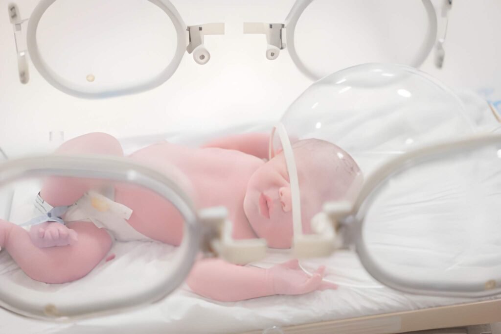
Diagnostic Pathways: Pulse Oximetry and Blood Gas
Modern neonatology relies on objective measurements to guide therapy. Pulse oximetry has become a universal screening tool in US hospitals, helping to catch "silent" hypoxemia that might not be visible to the naked eye. However, pulse oximetry only measures the percentage of hemoglobin saturated with oxygen; it does not reflect the total amount of oxygen in the blood or the carbon dioxide levels.
The ABG is the gold standard for diagnosing hypoxemia. It provides the partial pressure of oxygen (PaO2), the partial pressure of carbon dioxide (PaCO2), and the pH level. A PaO2 below 50 mmHg in a term newborn signifies significant hypoxemia. A low pH (acidosis) combined with low oxygen indicates that the lack of oxygen has forced the body to switch to anaerobic metabolism, producing lactic acid.
Critical Congenital Heart Disease (CCHD) screening involves placing a pulse oximeter on the right hand (pre-ductal) and either foot (post-ductal). A significant difference in saturation between these two sites suggests that blood is shunting away from the lungs, potentially indicating a major heart defect like transposition of the great arteries.
Management Protocols: Resuscitation and Beyond
The management of a newborn with low blood oxygen begins with the Neonatal Resuscitation Program (NRP) algorithm. The first step is providing warmth and stimulation. If the infant remains apneic or has a heart rate below 100, the clinician initiates Positive Pressure Ventilation (PPV). Interestingly, for term infants, the initial resuscitation usually begins with room air (21% oxygen) rather than 100% oxygen to avoid oxidative stress and tissue damage.
Oxygen Delivery Methods
If the infant needs ongoing support, clinicians choose from several delivery methods. Nasal cannulas provide low-flow oxygen, while Continuous Positive Airway Pressure (CPAP) keeps the lungs open during expiration. For infants with severe lung disease, mechanical ventilation becomes necessary. In cases of Persistent Pulmonary Hypertension of the Newborn (PPHN), inhaled nitric oxide—a potent vasodilator—is used to open the pulmonary arteries and improve blood flow to the lungs.
Clinicians calculate the "shunt fraction" to determine how much blood is bypassing the lungs entirely. This helps differentiate between lung disease (which responds well to oxygen) and heart disease (which may not).
PaO2 on 100% Oxygen: 150 mmHg
Normal Expected PaO2: 600 mmHg
Calculation: (600 - 150) / 20 = 22.5% Shunt
Outcome: A shunt greater than 20% indicates significant ventilation-perfusion mismatch or an anatomical heart defect.
Long-Term Outcomes and Neuroprotection
The long-term prognosis for a newborn with hypoxemia depends on the duration and severity of the deprivation. The brain is the most sensitive organ to oxygen deficits. If the deprivation is severe enough to cause Hypoxic-Ischemic Encephalopathy (HIE), the infant may face long-term developmental challenges, including cerebral palsy or cognitive delays.
A landmark advancement in neonatal care is therapeutic hypothermia, often called "cooling therapy." By lowering the infant's core body temperature to 33.5 degrees Celsius for 72 hours, clinicians can slow the metabolic rate and reduce the "secondary phase" of brain injury that occurs after oxygen is restored. This intervention has significantly improved the rates of survival without disability in infants affected by birth asphyxia.
Success in managing neonatal oxygen deficits requires a coordinated team of nurses, respiratory therapists, and neonatologists. With early detection through pulse oximetry and the application of modern neuroprotective strategies, the medical community continues to reduce the impact of birth-related hypoxemia. Providing a safe transition for every newborn remains the ultimate goal of perinatal medicine.


