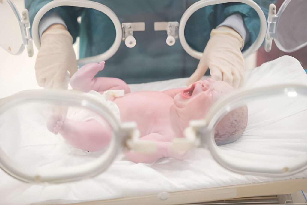
The presentation of a severely cyanotic newborn represents one of the most high-stakes emergencies in the neonatal nursery. Cyanosis, characterized by a bluish discoloration of the skin and mucous membranes, occurs when the absolute concentration of deoxygenated hemoglobin in the capillaries exceeds 5 grams per deciliter. While a mild bluish tint to the hands and feet (acrocyanosis) is normal during the first 24 to 48 hours of life, central cyanosis involving the tongue and trunk indicates a significant failure in oxygen delivery. Swift clinical differentiation between cardiac, respiratory, and metabolic causes is essential to ensure infant survival and neurological integrity.
Defining Central vs. Peripheral Cyanosis
Clinicians must immediately distinguish between central and peripheral presentations. Peripheral cyanosis, or acrocyanosis, usually results from cold exposure or peripheral vasoconstriction. In these cases, the oxygen saturation in the central circulation remains normal. Central cyanosis, however, signifies that the arterial blood itself is poorly oxygenated. The most reliable site to check for central cyanosis is the tongue and the mucous membranes of the mouth, as these areas have high vascularity and are less affected by ambient temperature.
When an infant appears blue, the medical team initiates a rapid evaluation of the "work of breathing." A severely cyanotic infant who is breathing comfortably (quiet tachypnea) is highly suspicious for a cardiac defect. Conversely, an infant with significant grunting, flaring, and retractions usually points toward a primary lung disorder. This observation forms the basis of the initial triage process.
The Triage of the Blue Newborn
The diagnostic pathway for severe cyanosis requires a systematic approach to rule out the most life-threatening conditions first. The medical team categorizes the presentation into four primary "buckets": Cardiac, Respiratory, Metabolic (Sepsis/Hypoglycemia), and Neurological (CNS depression).
| Observation | Cardiac Cause (CCHD) | Respiratory Cause (RDS/PPHN) |
|---|---|---|
| Respiratory Effort | Often "Comfortable" Tachypnea | Labored; Grunting and Retractions |
| Oxygen Response | Minimal to no improvement | Significant improvement with oxygen |
| Chest X-Ray | Clear lungs; abnormal heart shape | Opaque lungs; streaky or white-out |
| Cry Effect | Cyanosis worsens with crying | Cyanosis may improve with crying |
Critical Congenital Heart Disease (CCHD)
Severe cyanosis in a newborn is frequently a hallmark of "Critical Congenital Heart Disease." These are structural defects that prevent oxygenated blood from reaching the body's circulation. Many of these defects are "ductal dependent," meaning the infant relies on the fetal ductus arteriosus remaining open to survive. When the ductus begins to close naturally within the first hours or days of life, the infant's saturation levels plummet.
Pulmonary and Airway Failure
While cardiac issues are a primary concern, lung disorders are more common. Respiratory Distress Syndrome (RDS) is the leading cause of cyanosis in premature infants due to surfactant deficiency. In full-term infants, Persistent Pulmonary Hypertension of the Newborn (PPHN) is a frequent culprit. In PPHN, the blood vessels in the lungs remain constricted after birth, preventing blood from being oxygenated and forcing it to bypass the lungs through fetal shunts.
Meconium Aspiration Syndrome (MAS) can also cause severe cyanosis. If an infant inhales meconium (their first stool) into their lungs before or during birth, it creates chemical inflammation and mechanical blockage. This prevents effective gas exchange, leading to rapid desaturation. Pneumothorax, or a collapsed lung, is another acute emergency where air leaks into the chest cavity, compressing the lung and causing immediate, severe cyanosis.
The Hyperoxia Test Protocol
To differentiate between lung and heart issues, clinicians utilize the Hyperoxia Test. The infant is given 100 percent oxygen for 10 to 15 minutes. An arterial blood gas is then drawn to measure the partial pressure of oxygen (PaO2).
If the PaO2 rises above 150 or 200 mmHg, the cause is almost certainly respiratory. This indicates that the lungs simply needed more pressure or oxygen to function. If the PaO2 remains below 100 mmHg despite 100 percent oxygen, a cardiac shunt is likely. In structural heart disease, the blood physically cannot reach the lungs, so increasing the oxygen concentration does nothing to improve the saturation in the body.
Emergency Stabilization and Prostaglandin Use
When a newborn is severely cyanotic and a cardiac cause is suspected, the most critical intervention is the administration of Prostaglandin E1 (PGE1). This medication prevents the ductus arteriosus from closing or can even reopen a closed ductus. By keeping this fetal vessel open, the medical team ensures a "mixing" of blood or a pathway for blood to reach the lungs, buying time until surgery or further imaging can be performed.
Stabilization also involves careful temperature management and glucose monitoring. A cyanotic infant consumes energy at a massive rate, and hypoglycemia can quickly complicate the clinical picture. Intubation and mechanical ventilation may be necessary, especially if the cyanosis is respiratory or if the infant is showing signs of exhaustion. For PPHN, specialized treatments like Inhaled Nitric Oxide (iNO) can help relax the lung vessels and improve oxygenation.
Diagnostic Calculations in Cyanosis
Medical teams use various calculations to determine the severity of the oxygenation failure. One such calculation is the Oxygenation Index (OI), which helps decide if an infant needs advanced therapies like ECMO (heart-lung bypass).
Formula: (MAP x FiO2 x 100) / PaO2
Example Scenario:
Mean Airway Pressure: 15
FiO2 (Oxygen setting): 1.0 (100%)
Arterial PaO2: 50 mmHg
Calculation: (15 x 1.0 x 100) / 50 = 30
An OI greater than 25 indicates severe respiratory failure. An OI greater than 40 is a common threshold for considering ECMO therapy.
Surgical and Long-term Outlook
The long-term outlook for a cyanotic newborn depends entirely on the underlying cause. Many respiratory issues resolve within days or weeks with proper NICU support. Structural heart defects, while intimidating, are now largely repairable. Pediatric cardiothoracic surgeons can perform complex reconstructions on infants just days old. Early detection, often assisted by the Pulse Oximetry Screening performed on all newborns in the US, is the single most important factor in preventing sudden collapse and ensuring a healthy future.
The transition from a "blue baby" to a "pink baby" is a complex journey requiring the coordination of neonatologists, cardiologists, and respiratory therapists. For parents, understanding that cyanosis is a symptom of an underlying process—not the final diagnosis—is the first step in navigating the intensive care environment. With modern diagnostics and pharmacological interventions like prostaglandins, the vast majority of cyanotic newborns can be stabilized and successfully treated.


