Specialized Maternal Care
Pregnancy with Uterus Didelphys: Clinical Management of a Double Uterus
Table of Contents
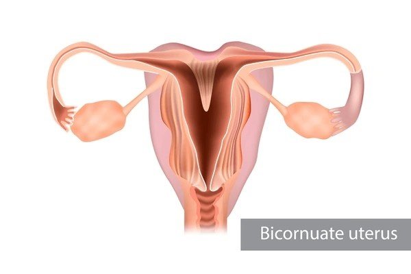
Defining Uterus Didelphys: An Anatomical Overview
Uterus didelphys, meaning "double uterus," is a rare congenital uterine anomaly affecting roughly $0.1$ percent of women. This condition arises during fetal development when the two paramesonephric (Mullerian) ducts fail to fuse completely along the midline. Instead of forming a single, unified uterus, two distinct uterine bodies and two cervices develop, often accompanied by a septum (wall) running vertically down the vagina, resulting in a double vaginal canal.
Crucially, a woman with Uterus Didelphys possesses two fully separate reproductive systems, each potentially capable of supporting a pregnancy. The two uteri, though connected by supportive ligaments, function autonomously, with independent cycles, blood supply, and, critically, different capacities for accommodating a growing fetus. Many women remain unaware they possess this anomaly until pregnancy or a routine gynecological examination.
Embryological Origins: The Unfused Ducts
Understanding the origin provides context for the risks. The Mullerian ducts usually fuse around the $10$th to $13$th week of embryonic development. The failure of this fusion results in two distinct systems. The impact on pregnancy stems from the fact that each resulting uterus is typically smaller and less muscular than a single, normal uterus, often leading to reduced distensibility—the ability to stretch and grow—and potentially compromised blood flow to the lining.
Pregnancy Scenarios in a Double Uterus
When a woman with Uterus Didelphys conceives, one of two primary scenarios occurs, each requiring different levels of vigilance and clinical action.
Pregnancy in One Uterus
The most common situation involves conception occurring in only one of the two uteri. The fertilized egg implants in one of the two cavities, and the pregnancy progresses there. The non-pregnant uterus continues its own hormonal cycle, though this is usually suppressed by the high levels of progesterone produced by the pregnant side.
A key observation in this scenario is the non-pregnant uterus may undergo a process called decidualization—the preparation of the lining—and may occasionally shed that lining, causing bleeding that mimics menstruation. This phenomenon can occur early in the pregnancy, leading to confusion, but is generally not harmful to the developing fetus in the adjacent uterus.
Twin Pregnancy (One Fetus in Each Uterus)
The rarest and most clinically complex scenario is a twin pregnancy where one fetus develops in each separate uterine cavity. This necessitates monitoring both uteri for growth, separate placental health, and the independent risk of rupture or preterm labor in each. It is also possible, though extremely rare, for a woman to conceive separately in each uterus weeks or even months apart (superfetation or superfecundation), leading to twins or even triplets with differing gestational ages.
The Clinical Risk Profile
While women with Uterus Didelphys are capable of carrying healthy pregnancies to term, the condition significantly elevates the risk for certain adverse outcomes. These pregnancies require the attention of a high-risk obstetrician or maternal-fetal medicine specialist.
Preterm Birth and Fetal Growth Restriction
Preterm delivery (birth before $37$ weeks gestation) is the most prominent concern. The reduced size and decreased muscular distensibility of the individual uterine cavity means the uterus may reach its maximum capacity sooner than a single, unified organ. This increased pressure can trigger preterm contractions and labor.
Furthermore, the atypical blood supply or smaller placental size within the compromised cavity increases the risk of Fetal Growth Restriction (FGR), where the fetus does not reach its full growth potential due to insufficient nutrition and oxygen supply. Monitoring fetal growth and uterine pressure is a key component of management.
Cervical Weakness and Miscarriage
The two cervices may also exhibit functional deficits. The condition is associated with a higher rate of second-trimester miscarriage due to cervical incompetence (a weak cervix that opens prematurely under the weight of the growing fetus). The non-pregnant cervix and uterus can also contribute to uterine irritability, increasing the risk of contractions.
Pregnancy Risk Comparison
| Adverse Outcome | Standard Risk (%) | Didelphys Risk (Approx. %) |
|---|---|---|
| Preterm Birth (before 37 weeks) | ~10% | 30% - 50% |
| Second Trimester Loss/Miscarriage | ~1% | 10% - 25% |
| Breech Presentation (at term) | ~3% | 20% - 30% |
This comparison highlights why clinical management must be adapted for high-risk monitoring, with a focus on mitigating preterm labor.
Specialized Management Strategies
Management of a Uterus Didelphys pregnancy involves focused interventions aimed at prolonging gestation and optimizing fetal position.
Progesterone Supplementation and Cerclage
Given the high risk of preterm birth, clinicians often implement preventative measures early in the second trimester:
- Cervical Length Monitoring: Weekly or bi-weekly transvaginal ultrasounds monitor the length of the pregnant cervix. Shortening indicates potential incompetence or preterm labor risk.
- Progesterone Therapy: Progesterone supplementation (often vaginal suppositories or intramuscular injections) is frequently used to relax the uterus and reduce the risk of spontaneous preterm birth.
- Cervical Cerclage: If the cervix shows significant shortening, a cerclage (a stitch placed around the cervix to keep it closed) may be placed to mechanically support the pregnancy until later gestation.
Delivery Planning and Presentation
The reduced uterine size affects the fetus's ability to rotate into the optimal head-down (vertex) position, leading to a higher incidence of non-vertex presentations.
Mode of Delivery
Approximately $80$ percent of pregnancies in a Uterus Didelphys result in a live birth, but the delivery mode is often affected by presentation.
- Increased Cesarean Rate: Due to the high rate of breech (feet or bottom first) or transverse (sideways) presentation, the Cesarean section rate is significantly higher than in the general population.
- Vaginal Birth Considerations: If the fetus is vertex (head down) and the pregnant uterus is unobstructed, a vaginal delivery is possible. However, the presence of the non-pregnant cervix or a vaginal septum may complicate the process, sometimes requiring surgical division of the septum prior to or during labor.
- Twinning Scenario: If twins are present (one in each uterus), a double Cesarean or a combination of vaginal and Cesarean delivery may be necessary, depending on the position of each fetus and the onset of labor in each uterus.
Interactive FAQ: Uterus Didelphys Management
Common questions regarding the management of a double uterus pregnancy. Click to reveal the expert answers.
The non-pregnant uterus does not actively interfere with the growth of the pregnant uterus. However, its physical presence can restrict the available space, limiting the pregnant uterus's capacity to stretch, which contributes to the higher risk of preterm labor and fetal growth restriction.
If you have a vaginal septum (a common feature of Uterus Didelphys), the decision depends on the planned delivery route. If a vaginal birth is planned, the septum may need surgical excision (metroplasty) before or during labor to allow for the fetus's passage. If a C-section is scheduled, the septum is typically left intact.
Not necessarily. Because the two uteri develop separately, they may have different sizes, blood supplies, and musculature. It is essential to treat a subsequent pregnancy, even if it is in the adjacent uterus, as a new high-risk scenario requiring the same level of close monitoring, especially for cervical length and preterm labor indicators.
Conclusion: Confident High-Risk Care
A pregnancy involving Uterus Didelphys is classified as high-risk, demanding a highly specialized approach to prenatal care. While the anatomical anomaly significantly increases the risk of preterm birth, miscarriage, and non-vertex presentations, the vast majority of these pregnancies result in successful live births under the care of a maternal-fetal medicine specialist. Consistent monitoring of cervical length, prophylactic progesterone use, and careful, pre-planned delivery strategies are the hallmarks of managing this condition, allowing expectant parents to proceed with informed confidence despite the anatomical challenges.


