The Second Ultrasound: The Comprehensive Fetal Anatomy Scan
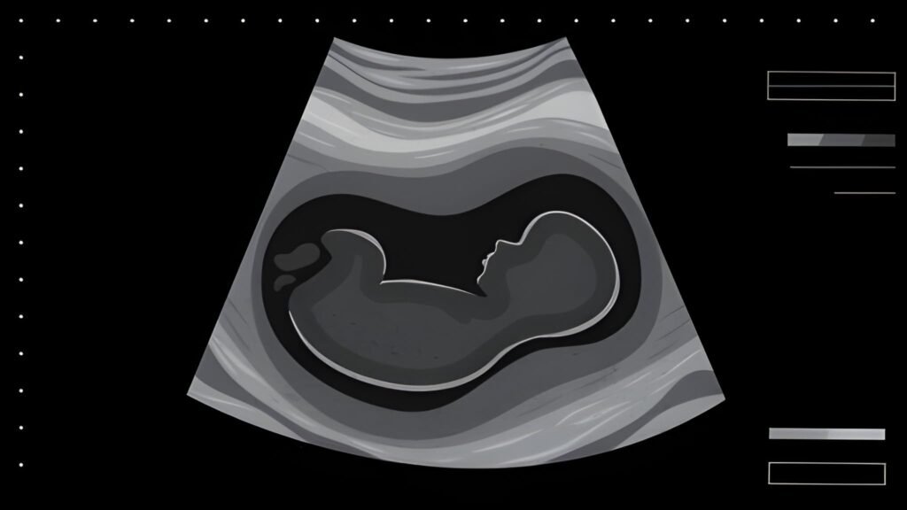
The second standard ultrasound, often called the **Anatomy Scan** or **Morphology Survey**, is the most comprehensive diagnostic imaging test of an uncomplicated pregnancy. Typically scheduled between 18 and 22 weeks, this detailed examination serves as the crucial clinical checkpoint of the mid-trimester. Unlike the first dating scan, which confirms viability and gestational age, the second ultrasound systematically investigates every major structure and organ system of the developing fetus.
As a specialist in maternal and child health, I view this scan as a partnership between technology and clinical expertise. It requires meticulous attention from the sonographer and patience from the expectant parents, as the examination can take anywhere from 30 minutes to over an hour. This guide outlines the key clinical objectives, the detailed procedures, and the specific findings that make this scan an indispensable part of prenatal care.
Table of Contents
1. Optimal Timing and Primary Purpose
The 18-to-22-week window is chosen precisely because the fetus is large enough for detailed visualization, but the ratio of amniotic fluid to baby size still allows for clear imaging of internal organs and tiny structures.
The Non-Negotiable Clinical Goal
The goal is to screen for **structural anomalies** (birth defects) that benefit from early diagnosis, allowing healthcare providers to plan specialized care immediately after birth. While the scan cannot detect all genetic conditions (like Down syndrome, which requires blood tests or amniocentesis), it identifies major structural issues, including approximately 90 percent of spinal defects and over 50 percent of congenital heart defects.
Scan Focus Shift: The Dating Scan focuses on confirming existence and due date. The Anatomy Scan focuses on **confirming integrity** and checking the architecture of every vital organ.
2. The Meticulous Anatomical Survey
The sonographer uses a systematic checklist to ensure every fetal system is properly evaluated. This process often requires the mother to shift positions to encourage the baby to move, optimizing the viewing angles.
Detailed Organ Systems Evaluation
The list below details the primary structures examined. Use the interactive section to learn why each system check is important:
The sonographer examines the cerebral ventricles, cerebellum, and the posterior fossa to assess overall brain development. The entire length of the spine is visualized in multiple planes to rule out **Spina Bifida**, and the skull integrity is checked.
This is a particularly time-intensive check. The sonographer must visualize the four chambers of the heart, confirm the two major vessels (aorta and pulmonary artery) exit correctly, and check the rhythm and rate of the heartbeat. This screens for congenital heart defects.
The scan confirms the presence of the stomach bubble (indicating the baby is swallowing amniotic fluid), the integrity of the abdominal wall, and the presence and function of both kidneys and the bladder. Kidney development is crucial, as the baby's urine production controls amniotic fluid volume.
The technician counts the long bones in the arms and legs (humerus, radius/ulna, femur, tibia/fibula) and examines the profile to rule out **cleft lip**. Fetal movement is observed to confirm neurological and muscular function.
3. Biometry: Checking Growth and Development
Growth measurements (biometry) confirm the baby is tracking appropriately for their gestational age. Since the due date is usually well-established by this point, these measurements primarily screen for growth anomalies.
Key Fetal Biometry Collected
| Measurement | Acronym | Clinical Purpose |
|---|---|---|
| Biparietal Diameter | BPD | Lateral width of the skull. |
| Head Circumference | HC | Total circumference of the skull. |
| Abdominal Circumference | AC | Circumference around the fetal abdomen (most sensitive to growth issues). |
| Femur Length | FL | Length of the thigh bone (used to confirm proper limb growth). |
4. Assessment of Maternal and Support Structures
The scan also provides essential information regarding the mother's anatomy and the support systems within the uterus.
Placental Location and Previa Risk
The location of the placenta and its relationship to the cervix (the exit from the uterus) is mapped. The primary concern is **Placenta Previa**, where the placenta lies low and covers the cervix. A previa diagnosis at 20 weeks is common, but most resolve as the uterus grows (apparent migration). If the placenta remains low, follow-up scans will be scheduled in the third trimester.
Amniotic Fluid Volume
The volume of amniotic fluid (measured as the Amniotic Fluid Index, or AFI) is calculated. Fluid volume must remain within a healthy range. Too little fluid (oligohydramnios) can signal problems with fetal kidneys, and too much fluid (polyhydramnios) may indicate gestational diabetes or fetal swallowing issues.
Cervical Length Check (for Preterm Risk)
Some providers may perform a Transvaginal Ultrasound (TVS) at the end of the anatomy scan to accurately measure the length of the cervix. This is particularly important for mothers with prior pregnancy complications or risk factors, as a short cervix is the strongest predictor of preterm birth risk.
5. Interpreting Results and Follow-up
The results of the anatomy scan are typically sent to your obstetrician for review. The sonographer is often prohibited from discussing clinical findings in detail.
Normal Scan and Fetal Gender
The vast majority of anatomy scans are reassuring, confirming the normal development of all structures. This is the stage where the gender of the baby is clearly visualized and can be disclosed if requested by the parents. Since the clinical work is complete, any additional imaging (3D or 4D photos) is usually performed at the end of the appointment.
Dealing with Atypical Results
If the sonographer suspects a structural anomaly or identifies a "soft marker" (a minor variation that increases the statistical risk for a chromosomal condition, such as a bright spot on the heart or dilated kidney pelvis), you will be immediately referred to a **Maternal-Fetal Medicine (MFM)** specialist. The MFM specialist provides high-level counseling and may order further diagnostic tests, including targeted Level 3 ultrasounds or amniocentesis, to achieve a definitive diagnosis.
The second ultrasound is a gift of insight and confirmation, providing essential architectural details of your developing child. Approach this appointment with patience and the confidence that early, informed diagnosis is the strongest tool available for ensuring the best possible outcome for the remainder of your pregnancy and delivery.


