I. The First Trimester: The Foundation (Weeks 1–13)
The first trimester, though often marked by invisible changes to the abdomen, represents the most rapid and critical phase of fetal development and hormonal adjustment for the mother. This stage spans from the first day of the last menstrual period (LMP) through the end of the 13th week.
Fetal Development: Organogenesis
During these 13 weeks, the fundamental structures of the entire human body are formed—a process called **organogenesis**. Within the first eight weeks, the embryo develops a beating heart, a neural tube (which becomes the brain and spinal cord), limbs, eyes, and ears. By the end of this trimester, the embryo transitions into a **fetus**, measuring about 3 inches long and weighing nearly one ounce. All major organs are in place, though they require continued development and refinement.
Maternal Experience: Hormonal Overload
The mother’s body is flooded with high levels of pregnancy hormones, primarily Progesterone and Human Chorionic Gonadotropin (hCG). These hormones trigger the hallmark symptoms of the first trimester:
- Profound Fatigue: The body is building the placenta, an energy-intensive process equivalent to running a marathon.
- Nausea and Vomiting: Often peaking around 9 weeks, commonly referred to as "morning sickness" (though it can occur anytime).
- Breast Tenderness: Hormones cause duct growth and increased blood flow, leading to swelling and heightened sensitivity.
Medical Milestones: The Intake and Viability Check
The primary medical focus is confirmation and dating. Key appointments and tests include:
- Initial Intake (8–12 Weeks): Comprehensive medical history, physical exam, and full blood panel (blood type, Rh factor, infectious disease screening).
- Dating USG: Confirms intrauterine location, detects a fetal heartbeat, and measures the Crown-Rump Length (CRL) to set the official Estimated Due Date (EDD).
- Genetic Screening Options: Review of non-invasive prenatal testing (NIPT) and Nuchal Translucency (NT) scan options.
II. The Second Trimester: The Golden Period (Weeks 14–27)
The second trimester is often called the "Golden Period" of pregnancy. It spans from the start of the 14th week through the end of the 27th week. Symptoms from the first trimester typically subside, and energy levels return, making this the most comfortable phase for many expectant mothers.
Fetal Development: Growth and Refinement
The risk of miscarriage drops significantly after the first trimester. Fetal development shifts from forming structures to rapidly growing and refining organ systems. The fetus grows from roughly 3 inches to nearly 14 inches long and weighs about 2 pounds by the end of the 27th week. Milestones include:
- Quickening (16–22 Weeks): The mother feels the first subtle fetal movements.
- Hearing Development: The fetus begins to hear muffled external sounds and the distinct sound of the mother’s voice and heartbeat.
- Lung Maturation: Lungs begin developing their surfactant cells, although they are not yet mature enough for survival outside the womb until the end of this trimester.
Maternal Experience: Visible Changes and New Comfort
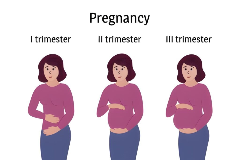
Physical changes become visibly apparent. The uterus rises out of the pelvis and into the abdominal cavity, requiring maternity wear. New symptoms replace the old:
- Increased Energy: Fatigue and nausea typically resolve, and appetite increases.
- Physical Aches: Ligaments stretch (Round Ligament Pain), leading to lower back and pelvic discomfort.
- Skin Changes: The linea nigra (dark line down the abdomen) and facial darkening (melasma) may appear due to hormonal pigmentation changes.
Medical Milestones: Anatomy and Screening
The focus is placed on detailed structural assessment and growth monitoring:
- Anatomy Scan (18–22 Weeks): The most detailed USG, checking the structure of all major organs, limbs, spine, and brain. The fetus’s sex is usually discernible at this time.
- Glucose Screening (24–28 Weeks): Testing for Gestational Diabetes Mellitus (GDM), a temporary form of diabetes common in late pregnancy.
- Amniocentesis: An invasive diagnostic test offered to high-risk patients to confirm genetic anomalies.
- Routine Appointments: Frequency shifts to monthly checks of weight, blood pressure, and fundal height.
III. The Third Trimester: Preparation and Completion (Weeks 28–40)
The third trimester, extending from the 28th week until delivery, is dedicated to final fetal growth, organ maturation, and the mother’s physical and psychological preparation for labor and birth. It is often the most physically demanding stage.
Fetal Development: Weight Gain and Maturation
The fetus gains the majority of its body fat during this time, preparing for temperature regulation outside the womb. Key developmental processes include:
- Lung Maturity: The lungs complete maturation, improving survival chances dramatically after 37 weeks.
- Brain Growth: The brain undergoes rapid expansion, tripling in weight during this final trimester.
- Fetal Movement: Movements become powerful, though less frequent towards the end as space runs out. Fetal monitoring is crucial to ensure continued well-being.
Maternal Experience: Physical Stress and Anticipation
Discomfort intensifies as the uterus places extreme pressure on internal organs and the diaphragm. Common symptoms include:
- Shortness of Breath: The uterus pushes up against the lungs, reducing lung capacity.
- Frequent Urination: The baby's head drops down into the pelvis (lightening), putting direct pressure on the bladder.
- Swelling (Edema): Fluid retention often causes swelling in the feet, ankles, and hands.
- Braxton Hicks Contractions: The uterus practices for labor with these irregular, painless tightening sensations.
Medical Milestones: Monitoring and Delivery Prep
Appointments increase in frequency, reflecting the higher risks associated with the final weeks:
| Weeks | Routine Activity | Key Tests Performed |
|---|---|---|
| 28 – 36 Weeks | Appointments every 2 weeks. Fundal height checks, fetal movement counts. | Rh immunoglobulin (Rhogam) injection if Rh-negative; Tdap vaccine if not recent. |
| 36 – 40 Weeks | Weekly appointments. | Group B Streptococcus (GBS) swab test; cervical checks (if agreed upon); discussion of birth plan. |
| 41 Weeks + | Non-Stress Tests (NSTs) and Biophysical Profiles (BPPs) to assess placental function. | Induction discussions to prevent post-term risks. |
Fetal Development Summary: Size Progression
The speed of development across the three trimesters is astonishing, moving from a microscopic cell cluster to a full-term infant capable of independent life.
- End of First Trimester (13 Weeks): Length of a peach (about 3 inches). Weight of a lemon (about 1 ounce).
- End of Second Trimester (27 Weeks): Length of a head of lettuce (about 14 inches). Weight of a large turnip (about 2 pounds).
- End of Third Trimester (40 Weeks): Length of a small watermelon (19–21 inches). Weight of a cantaloupe (6–9 pounds).
The Fourth Trimester: Postpartum Recovery and Adjustment
While pregnancy officially ends at birth, the concept of the **Fourth Trimester**—the 12 weeks following delivery—is now widely recognized in maternal and child health. This phase is critical for the mother’s physical healing and the newborn’s adjustment to life outside the womb.
Maternal Recovery and Monitoring
The body undergoes rapid and intense changes as it returns to the non-pregnant state. Key components of the fourth trimester:
- Uterine Involution: The uterus rapidly shrinks from the size of a watermelon back to its pre-pregnancy size.
- Hormone Shift: Progesterone and estrogen levels drop dramatically, triggering milk production and often contributing to postpartum mood shifts.
- Physical Healing: Recovery from vaginal birth tears or Cesarean section incisions. The traditional six-week checkup is now often supplemented by earlier and later appointments to address long-term health and mood disorders.
Newborn Adjustment and Attachment
For the child, the fourth trimester is defined by external adaptation, establishing feeding routines, and rapid neurodevelopment. The goal is to provide a calm, womb-like environment to aid this transition, emphasizing skin-to-skin contact and responsive feeding.


