Advanced Fetal Imaging
3D Sonography: Clinical Insight, Fetal Bonding, and Safety Guidelines
Table of Contents
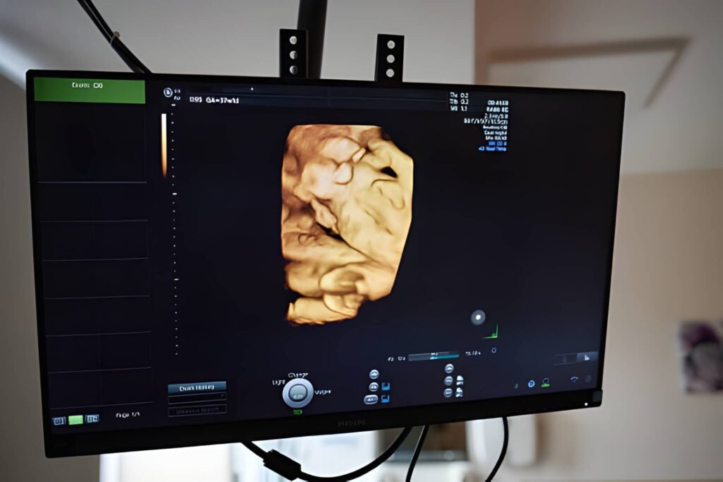
Defining 3D Sonography: More Than Just a Photo
Three-dimensional (3D) sonography is a sophisticated imaging technology that has revolutionized fetal assessment. Unlike the traditional two-dimensional (2D) ultrasound, which produces flat, grayscale, cross-sectional slices of the fetus, 3D ultrasound utilizes software to generate a lifelike volume of data. This volume can then be manipulated, viewed from multiple angles, and rendered to show surface features of the fetus, providing depth perception that is impossible to achieve with 2D alone.
While often sought for aesthetic reasons—offering parents their first clear glimpse of their baby's face—its true value lies in its clinical ability to confirm the presence or absence of specific structural anomalies. The use of 3D sonography is a powerful example of technology enhancing both medical accuracy and the parental experience of gestation.
The Science of Volume Rendering
The fundamental physics of ultrasound remains the same in 2D and 3D imaging: sound waves travel into the body, reflect off tissues, and return to a transducer. The difference lies in data acquisition and processing.
Collecting the Volume Data
A 3D transducer is designed to sweep through an area, collecting hundreds of 2D images in rapid succession. These slices are collected to form a cube of information, known as the volume data set. The imaging machine then uses specialized computer algorithms (volume rendering) to assemble these slices into a unified, volumetric image.
This process allows the clinician to examine the fetus not just by slicing through it, but by looking at surfaces and planes simultaneously. For example, a 2D scan might show a potential gap in the lip, but a 3D scan provides a complete topographical view of the fetal face, confirming the extent and severity of a potential cleft palate in a single image.
Clinical Value: Diagnosis Beyond 2D
While 2D ultrasound remains the backbone of obstetric screening, 3D sonography is utilized by maternal-fetal medicine (MFM) specialists when structural anomalies are suspected or require definitive clarification.
Refining Fetal Anomaly Diagnosis
The clearest clinical benefit of 3D imaging lies in evaluating the surface anatomy of the fetus.
- Cleft Lip and Palate: 3D provides a superior visual confirmation of facial structure, which is crucial for multidisciplinary surgical planning before birth.
- Skeletal Abnormalities: It helps visualize the exact alignment and condition of bones, especially in conditions affecting the limbs or spine (like spina bifida).
- Placental Visualization: 3D imaging enhances the evaluation of the placenta's structure and attachment, particularly in cases of placenta previa or suspected placenta accreta (where the placenta attaches too deeply into the uterine wall), which are high-risk conditions requiring specialized delivery planning.
Relevance for Prenatal Counseling
In the socioeconomic landscape of the United States, obtaining a definitive diagnosis is critical. When a structural anomaly is confirmed via 3D imaging, it allows parents to engage with specialized pediatric surgeons and counselors well before delivery. This preparation—logistically and emotionally—is essential for ensuring seamless, immediate care for the newborn and mitigating the financial stress associated with unexpected, complex medical needs after birth.
Optimal Timing: When to Schedule a 3D Scan
The clarity and quality of the 3D image depend significantly on the amount of amniotic fluid present and the baby's position.
The Best Imaging Window
The ideal window for capturing clear, high-quality 3D images is typically between **26 and 32 weeks** gestation.
- Before 26 Weeks: The baby has very little subcutaneous fat, making the face look skeletal and less recognizable.
- After 32 Weeks: The baby has grown considerably, and the space (amniotic fluid) around the face diminishes, making it difficult to obtain a clear, unobstructed image, as the baby's face may be pressed against the uterine wall or the placenta.
Interactive: 2D, 3D, and 4D Distinction
The world of advanced sonography uses confusing terms. Use the tabs below to clarify the differences between the common imaging types.
2D Ultrasound: The Clinical Standard
- Output: Flat, grayscale, cross-sectional slices.
- Purpose: Primary tool for measuring anatomy (biometry), assessing internal organs, confirming viability, and tracking fetal growth.
- Timeline: Used throughout the entire pregnancy, most commonly for the anatomy scan (18–22 weeks).
3D Sonography: Static Volume
- Output: A still, three-dimensional image showing the surface features of the fetus (like a photo).
- Purpose: Used for specialized confirmation of external structural anomalies (face, limbs) and for parental bonding.
- Benefit: Provides depth perception for clinical confirmation of surfaces.
4D Ultrasound: 3D in Motion
- Output: A live, moving video feed of the 3D volume (the fourth dimension is time).
- Purpose: Allows parents and clinicians to observe fetal behavior in real-time (yawning, sucking thumb, smiling).
- Benefit: Primarily utilized for enhanced parent-child bonding and assessing fetal neurological development.
Safety Standards and Professional Use
The safety profile of 3D sonography is identical to that of traditional 2D ultrasound, as both use the same non-ionizing, mechanical energy (sound waves). However, professional regulatory bodies emphasize guidelines regarding unnecessary exposure.
FDA and Professional Recommendations
The US Food and Drug Administration (FDA) and the American Institute of Ultrasound in Medicine (AIUM) recommend that ultrasound scans—including 3D/4D—should only be performed by trained professionals for **medical necessity** and kept to the lowest practical power setting and shortest duration necessary to achieve the diagnostic goal.
This guidance strongly discourages non-medical, commercial "keepsake" scans performed solely for aesthetic pleasure. While the sound waves are safe, prolonged exposure at high settings (sometimes used for better aesthetic results) increases the baby's thermal exposure, which may pose a theoretical risk. Parents should always ensure 3D scans are performed in a clinically certified setting, ideally integrated into their established prenatal care plan.
Emotional Benefit: Fetal Bonding and Parenthood
Beyond the clinical charts and measurements, 3D sonography plays a crucial role in maternal and paternal fetal bonding.
For many expectant parents, seeing the recognizable features of their baby—a defined nose, chin, or the gesture of a hand—transforms the abstract concept of pregnancy into a concrete reality. This visual connection is often critical for fathers and partners who do not experience the physical reality of gestation. Strong fetal bonding, initiated by clear 3D images, positively correlates with parental attachment post-delivery and enhances the psychological confidence of new parents. In this way, 3D sonography serves a vital socioeconomic purpose by strengthening the family unit's foundation even before the birth event.
Conclusion: Integrating Technology and Care
3D sonography stands as a vital tool in modern obstetrics, leveraging volume rendering to provide depth and clarity unmatched by 2D imaging. Its primary benefits are clinical—refining the diagnosis of structural anomalies and guiding complex planning—but its role in enhancing fetal bonding is equally significant. When performed within the optimal 26-to-32-week window by qualified professionals and adhering strictly to safety protocols, 3D and 4D imaging confidently integrate technological advancement with compassionate prenatal care, preparing both the medical team and the family for the journey ahead.


