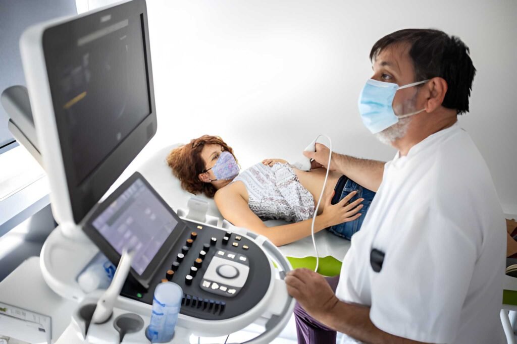
Equine Gestation: Clinical Findings of the 60-Day Pregnancy Ultrasound
The 60-day mark (approximately nine weeks) represents the conclusion of the most common risk period for early embryonic loss (EEL) in the mare. The initial, highly mobile embryonic phase is long past, and the pregnancy is now established in the uterine horn. The primary purpose of the ultrasound at this stage—performed via the **transrectal approach**—is to provide the final confirmation of viability before the conceptus grows too large for detailed visualization, and to conduct the initial fetal sexing procedure.
As a specialist in reproductive health, I confirm that successful management of the mare hinges on this detailed assessment. The 60-day scan shifts the focus from simple growth rates to the physical development of the fetus and the hormonal support provided by the mother.
Table of Contents
1. Fetal Size and Visual Confirmation
By 60 days gestation, the embryonic vesicle (the fluid-filled sac) has grown substantially. The uterus containing the conceptus is now large, measuring roughly 12 centimeters in diameter, filling a large portion of the uterine horn.
Milestones Visible at Day 60
- Organogenesis Complete: All major organs and body systems are fully formed and functioning. The conceptus is now accurately referred to as a **fetus**.
- Recognizable Form: The fetus is fully recognizable as an equine conceptus, with visible head, backbone, chest, and limb buds. The focus of the fetus is now dedicated to maturation and growth over the remaining months.
- Fetal Heart Rate: The heartbeat, first visible around Day 24, should be strong and easily detectable.
| Gestation Day (from Ovulation) | Vesicle Diameter (Approx.) | Key Event on Ultrasound |
|---|---|---|
| Day 14 | 13-18 mm | Pregnancy confirmed; Twins identified. |
| Day 28 | 3-4 cm | Heartbeat definitively detected. |
| Day 60 | 12 cm | Fetus visible, Ossification occurring, Sexing possible. |
2. Hormonal and Placental Support Structures
The 60-day scan provides a critical check of the support structures that ensure the continuation of the pregnancy beyond the 100-day mark. The early hormonal system established in the first month is now backed by a specialized system.
Endometrial Cups and eCG Production
Between days 35 and 120, specialized structures called **endometrial cups** form within the mare's uterus. These cups produce **equine Chorionic Gonadotropin (eCG)**. This hormone stimulates the ovaries to form accessory Corpora Lutea (CLs), which secrete large amounts of progesterone. The 60-day scan confirms the proper size and condition of the accessory CLs and reviews the functionality of the utero-placental interface, ensuring adequate progesterone support is in place for the second trimester.
3. The Fetal Sexing Window: Procedure and Timing
The 60-day scan is often the earliest time fetal sexing can be performed accurately. This is an elective procedure but is valuable for breeding management and sales purposes.
Ultrasonographic Method for Sex Determination
Sex determination is possible because the internal genital bud, or **genital tubercle**, migrates to a distinct location based on sex:
Male (Colt): The genital tubercle migrates forward, positioning itself between the fetus’s hind limbs.
Female (Filly): The genital tubercle migrates backward, positioning itself underneath the tail.
The veterinarian can identify the location of this structure with a transrectal ultrasound probe. If the fetus is positioned poorly, the procedure may be postponed until the next optimal window (around Day 120-150) or until movement allows better visualization.
4. Monitoring Growth and Fetal Movement
Although detailed anatomical checks were performed earlier, the scan at 60 days provides baseline measurements that can be used to assess late-stage growth rates, particularly in high-risk mares.
Fetal Activity and Wellbeing
The fetus is now highly active within the allantoic sac, making frequent limb movements, head nods, and body shifts. The ultrasound confirms these vigorous movements. The lack of visible, predictable movement may raise concerns about fetal distress or viability, prompting closer monitoring.
5. Management Decisions Post-60 Days
Successful completion of the 60-day scan drastically reduces the clinical need for frequent subsequent intervention. The veterinarian's focus moves to preventative health care and preparing for the final trimester.
Post-60 Day Care Checklist
- Vaccination Schedule: Review the vaccination plan. Often, a rhino (Equine Herpesvirus, EHV-1) vaccination series begins around the fifth, seventh, and ninth months of gestation to prevent viral abortion.
- Nutritional Plan: Although major caloric increases are reserved for the final trimester, ensure the mare maintains a moderate to good body condition score. Obesity or poor body condition can complicate delivery.
- Future Scans: Unless the mare is high-risk, subsequent scans may be elective for owners or scheduled only around Day 90–120 for the second sexing window.
The 60-day ultrasound confirms that the physiological risks of early loss are largely past and provides the final opportunity for critical diagnostic procedures like sexing. By confirming the well-being and managing the environment and nutrition, you secure the foundation for a healthy gestation leading toward foaling.


