The 20-Week Appointment: Anatomy Scan and Halfway Checkup
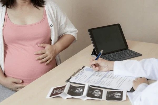
Reaching 20 weeks marks the midpoint of pregnancy—a significant milestone known as the halfway mark. The corresponding prenatal appointment is perhaps the most anticipated and diagnostically crucial visit. Its centerpiece is the **Anatomy Scan** (or Fetal Morphology Survey), a detailed, non-invasive ultrasound that systematically checks every major structure of the developing baby. This appointment shifts the focus from simple growth to comprehensive health assurance, providing invaluable data for the rest of your pregnancy.
As a specialist in child and mother health, I guide expectant parents through the dual components of this visit: the routine maternal health checks and the in-depth fetal examination. Understanding the clinical objectives of each step transforms this long appointment from a simple viewing into a powerful tool for informed prenatal care.
Table of Contents
1. The Routine Maternal Health Assessment
Before or after the lengthy ultrasound, your healthcare provider will perform the standard checks that are a feature of every prenatal visit. These ensure your body is managing the physiological demands of the second trimester efficiently.
Maternal Vitals and Weight Management
- Weight and Blood Pressure: Consistent monitoring ensures healthy weight gain and screens for potential issues like gestational hypertension or preeclampsia, though preeclampsia is typically a late-trimester concern.
- Urine Dipstick: A quick check for glucose (signaling potential gestational diabetes) and protein (another potential early marker for preeclampsia or kidney issues) and signs of infection.
- Fetal Heart Rate: The Doppler is used to listen to the baby's heart rate, confirming general well-being and viability before the more detailed ultrasound scan.
- Fundal Height: The provider begins measuring the fundal height (the distance from the pubic bone to the top of the uterus). At 20 weeks, the top of the uterus usually aligns with the navel (umbilicus).
Fetal Movement and Quickening
The 20-week mark is often when expectant mothers begin to feel **quickening**—the first subtle flutters of fetal movement. Your provider will inquire about feeling these movements. If you have not felt anything yet, it is generally not a concern, especially for first-time mothers. However, consistent movement is a vital sign of fetal well-being moving forward.
2. The Anatomy Scan: A Systematic Fetal Checkup
The anatomy scan is a comprehensive, non-stressful examination that typically lasts between 30 and 60 minutes. The sonographer systematically evaluates the baby's entire structure, ensuring development aligns with the gestational age. The goal is to detect approximately 50 to 90 percent of all major congenital structural anomalies, providing critical time for planning specialized care if needed.
The Detailed Anatomical Checklist
The sonographer must obtain clear images of all major systems. If the baby is uncooperative (or "camera shy"), you may be asked to walk around, use the restroom, or drink a cold beverage to encourage movement.
Interactive Checklist of Major Systems Examined
- Brain Structure: Verification of the cerebellum, corpus callosum, and cerebral ventricles to rule out structural anomalies like hydrocephalus.
- Spine: Detailed view of the vertebrae in both longitudinal and transverse planes to screen for neural tube defects, particularly spina bifida.
- Face: Examination of the orbits (eye sockets), nasal bone, and lips to check for conditions like cleft lip or palate.
- The Four-Chamber Heart View: A critical check ensuring the four chambers and two major outflow tracts (aorta and pulmonary artery) are correctly formed and functioning.
- Lungs and Diaphragm: Examination of the chest cavity and the diaphragm, the muscle separating the chest from the abdomen.
- Kidneys and Bladder: Confirming the presence of both kidneys, checking for internal dilation, and ensuring the bladder fills and empties (a sign the urinary system is functional).
- Stomach: Visualization of the stomach bubble confirms the baby is swallowing amniotic fluid.
- Extremities: Verification of the presence and proper formation of all four limbs, including fingers and toes (often difficult to count definitively).
- Umbilical Cord: Checking the cord for the correct number of vessels (two arteries and one vein) and ensuring proper insertion into the fetal abdomen and placenta.
- Sex: If requested and visible, the gender of the baby is identified at this stage.
3. Biometry: Key Measurements of Growth
The sonographer captures standardized measurements (biometry) to calculate the estimated fetal weight (EFW) and confirm that the baby’s growth rate aligns with the established due date.
Biometric Data Collected at 20 Weeks
These measurements help screen for Fetal Growth Restriction (FGR) or excessive growth that might signal gestational diabetes later in pregnancy:
| Measurement | Acronym | Clinical Purpose |
|---|---|---|
| Biparietal Diameter | BPD | Width of the head, used to assess head shape and size. |
| Head Circumference | HC | Overall head size, crucial for brain growth tracking. |
| Abdominal Circumference | AC | Measurement around the baby's tummy, often the first indicator of growth issues or FGR. |
| Femur Length | FL | Length of the thigh bone, used to assess long bone development. |
4. Placental Position and Cervical Length
The appointment also involves a crucial evaluation of the maternal structures supporting the pregnancy.
Placenta Position and the Previa Concern
The location of the placenta is critical. The scan must check for **placenta previa**, a condition where the placenta covers part or all of the cervix. While a low-lying placenta at 20 weeks is common, the provider needs to track its position. As the uterus expands, the placenta often migrates away from the cervix (a process called apparent migration). If the placenta remains low in the third trimester, it requires specialized management and often a Cesarean delivery.
Cervical Length Assessment
In many clinics, a transvaginal ultrasound (TVS) is offered or performed at the end of the anatomy scan to accurately measure the length of the cervix. This measurement is a key predictor of preterm labor risk. A short cervix may necessitate preventative interventions like progesterone supplementation or a cerclage.
5. Preparing for the Results and Next Steps
The anatomy scan is a comprehensive screening tool, not a perfect diagnostic test. It finds structural issues, but it cannot rule out chromosomal or genetic conditions entirely.
Interpreting the Results
- Normal Findings: Most scans yield reassuring results. The sonographer may inform you immediately that all structures appear normal, though the official report must be reviewed by the maternal-fetal medicine specialist or your OB-GYN.
- Atypical Findings: If a soft marker (a subtle finding that slightly increases risk for a chromosomal issue) or a potential structural anomaly is detected, you will be referred for immediate consultation with a specialist for further diagnostic testing, such as amniocentesis or a targeted Level 3 ultrasound.
Upcoming Appointments and Glucose Screening
The 20-week appointment often involves planning for the next major clinical screen: the **Glucose Tolerance Test (GTT)**, typically scheduled between 24 and 28 weeks to check for gestational diabetes. Your provider will often give you instructions or the sugary drink required for the test during this visit.
The 20-week appointment is a pivotal moment that provides a detailed, non-invasive glimpse into your baby's current health and development. Approach this visit informed, allowing the detailed scan to provide the clinical certainty needed for a confident transition into the second half of your pregnancy.


