Mid-Pregnancy Diagnostic Imaging
The Level 2 Ultrasound: A Detailed Guide to the Pregnancy Anatomy Scan
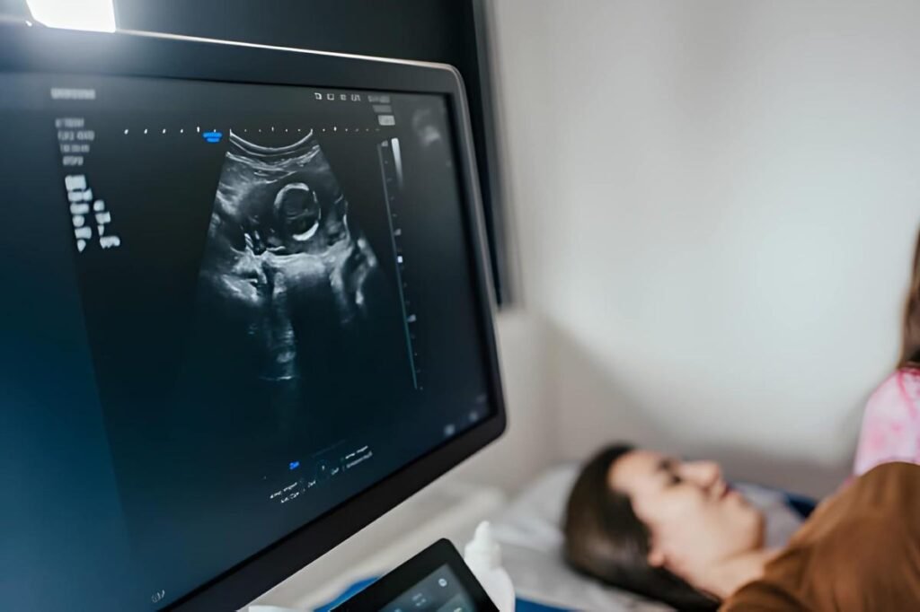
The Level 2 Ultrasound, universally known as the Anatomy Scan, is the most comprehensive fetal assessment performed during pregnancy. Scheduled typically between 18 and 22 weeks, this detailed imaging appointment shifts the focus from simple viability and dating to the meticulous examination of every organ, bone, and system within the developing fetus. It is a critical diagnostic tool used to confirm normal development, identify potential structural anomalies, and assess the supportive structures of the pregnancy (placenta, umbilical cord, amniotic fluid). Approaching this highly detailed scan with clarity ensures a focused and informed experience.
Table of Contents
Optimal Timing and Purpose
The 18-to-22-week window is optimal because the fetus is large enough for detailed visualization of organ systems, yet there remains sufficient amniotic fluid to create clear acoustic windows for the sound waves.
The Primary Objectives of the Scan
- Structural Assessment: To identify major congenital anomalies that require specialized management during pregnancy or immediately after birth.
- Growth Confirmation: To verify that the fetus is growing at an appropriate pace, aligning with the expected gestational age.
- Placental Location: To determine where the placenta attaches to the uterine wall, assessing for conditions like placenta previa.
- Gender Determination: To identify the biological sex of the fetus, if desired and if the anatomy is clearly visible.
Level 2 vs. Targeted Ultrasound
The term "Level 2 Ultrasound" is often used interchangeably with "Anatomy Scan" for a high-detail, routine mid-pregnancy scan. However, a "Targeted" or "Comprehensive" scan may be ordered specifically for high-risk pregnancies, such as those with a history of congenital defects or high-risk genetic screening results. This scan is generally performed by a maternal-fetal medicine (MFM) specialist and focuses increased attention on specific areas of concern.
Meticulous Fetal Anatomy Checklist
The anatomy scan involves a systematic review of the entire body, ensuring the sonographer captures images and measurements of over a dozen critical fetal structures. The examination is methodical, often requiring the sonographer to wait patiently for the fetus to move into specific positions.
Organ Systems Under Review
- The Brain: Examination includes the cerebellum, choroid plexus, lateral ventricles, and cavum septum pellucidum. The provider looks for correct development and size of intracranial structures, ruling out conditions like hydrocephalus.
- The Face and Neck: Assessment covers the profile, upper lip, and nostrils to check for cleft lip or palate. The orbits (eye sockets) are also measured.
- The Heart: This is a highly critical part of the scan. The sonographer assesses the four chambers of the heart, the proper formation of the outflow tracts (aortic and pulmonary arteries), and the rhythm of the heartbeat.
- The Abdomen and Chest: The stomach bubble, liver, kidneys (for hydronephrosis), and bladder are identified. The provider confirms the abdominal wall is completely closed and that the bowel is properly contained.
- The Spine and Limbs: The entire length of the spine is viewed in both longitudinal and transverse planes to exclude neural tube defects like spina bifida. All four limbs are visualized, confirming the presence of hands, feet, and the correct number of digits.
Assessment of Supportive Structures
The health of the supporting structures is as vital as the fetal anatomy itself. These elements determine the safety and longevity of the pregnancy.
Placenta, Cord, and Fluid
- Placental Location: The distance between the lower edge of the placenta and the internal cervical os (opening of the uterus) is measured. If the placenta covers the os, Placenta Previa is diagnosed. An early diagnosis allows the care team to prepare for potential bleeding and plan for a C-section if the condition persists.
- Amniotic Fluid Volume: Measured using the Amniotic Fluid Index (AFI). Too little fluid (oligohydramnios) or too much fluid (polyhydramnios) can signal fetal kidney problems, growth restriction, or other issues.
- Umbilical Cord: The cord is visualized to confirm the presence of three vessels (two arteries and one vein). A two-vessel cord (Single Umbilical Artery or SUA) often warrants extra screening.
Fetal Growth and Biometry Measurements
The Level 2 scan confirms or adjusts the due date, comparing the fetal biometry (measurements) to expected norms. The measurements are critical for predicting fetal weight and identifying growth restriction.
Key Biometry Measurements
| Measurement Abbreviation | Definition | Purpose in the Anatomy Scan |
|---|---|---|
| BPD | Biparietal Diameter (across the head) | Assess head size and brain development. |
| HC | Head Circumference | A secondary check on head size and dating accuracy. |
| AC | Abdominal Circumference | Primary measure for monitoring fetal nutrition and growth (Macrosomia/FGR). |
| FL | Femur Length (thigh bone) | Assess long bone growth and confirm dating. |
Interpreting Anomalies and Soft Markers
The vast majority of anatomy scans show normal development. However, the scan sometimes detects findings categorized as either major anomalies or "soft markers."
Distinguishing Between Findings
- Major Structural Anomaly: A defect requiring surgical correction after birth or specialized prenatal monitoring (e.g., severe heart defect, kidney agenesis, spina bifida). These findings often require referral to an MFM specialist and genetic counseling.
- Soft Marker: A minor anatomical variant or transient finding that does not usually cause health problems on its own but is associated with a slightly increased risk of a chromosomal condition (e.g., Trisomy 21). Common soft markers include an Echogenic Intracardiac Focus (EIF) or Pyelectasis (mild kidney dilation).
Interactive Tool: Understanding the Impact of Soft Markers
Soft Marker Risk Check
If a soft marker was found, understand that the risk changes based on other factors.
Practical Preparation for the Appointment
A small amount of proactive preparation helps ensure the scan is successful and that you get the most out of the two-hour experience.
Tips for Clearer Imaging
- Hydration: Drink plenty of water in the days leading up to the appointment. Good hydration improves amniotic fluid clarity and transmission of the ultrasound waves.
- Bladder: Unlike the very first scan, a full bladder is usually NOT required for the mid-pregnancy anatomy scan (abdominal approach). However, check with your clinic for specific instructions.
- Fetal Positioning: Some providers suggest consuming a small amount of juice or a light snack 15 minutes before the scan to encourage the fetus to move away from a difficult position.
The Level 2 Ultrasound is a detailed, reassuring checkpoint that provides a complete anatomical roadmap of the developing fetus. By understanding the breadth of the examination, you transition from a general state of anticipation to a state of informed certainty, ready to partner with your care team in managing the rest of the pregnancy journey with confidence.


