Clinical Management of Multisite Bleeding in the Neonate
Navigating Melena, Umbilical Hemorrhage, and Hematuria in the First Week of Life
Contents
Watching a newborn experience bleeding from multiple sites is one of the most distressing scenarios for both parents and healthcare providers. When a neonate presents with melena (dark, tarry stools), umbilical bleeding, and hematuria (blood in the urine), the clinical picture points toward a systemic coagulation failure rather than a localized injury. Prompt recognition and aggressive intervention are paramount to preventing long-term neurological damage or hypovolemic shock.
The Clinical Presentation: Identifying Bleeding Sites
In the neonatal period, "visible" bleeding is often the tip of the iceberg. Multisite bleeding indicates that the infant's natural clotting mechanisms are severely compromised. Let’s break down the three primary presentations mentioned:
Melena
Melena refers to black, sticky, tarry stools. This color indicates that blood has been partially digested, suggesting an upper gastrointestinal source or significant ingestion of blood. In newborns, it is a hallmark sign of internal gastrointestinal hemorrhage.
Umbilical Oozing
While minor oozing can happen when the cord separates, persistent or heavy bleeding from the umbilical stump is a major red flag. It is often the first "external" sign of a systemic bleeding disorder.
Hematuria
Blood in the urine (gross hematuria) is relatively rare in healthy newborns. When it occurs alongside other bleeding, it suggests renal involvement or a profound systemic inability to maintain vascular integrity.
Primary Etiologies: Why Is the Baby Bleeding?
The differential diagnosis for a bleeding neonate is narrow but critical. Most cases fall into one of three categories: Vitamin K deficiency, consumption of clotting factors (sepsis/DIC), or inherited factor deficiencies.
1. Vitamin K Deficiency Bleeding (VKDB)
This is the most common cause of spontaneous bleeding in an otherwise healthy-appearing newborn. Because Vitamin K does not cross the placenta well and breast milk contains low levels, infants are born with a natural deficiency. Without a prophylactic injection at birth, the liver cannot produce active clotting factors II, VII, IX, and X.
2. Disseminated Intravascular Coagulation (DIC)
Unlike VKDB, infants with DIC usually look "sick." They may have a history of birth asphyxia, respiratory distress, or maternal infection. In DIC, the body uses up all its platelets and clotting factors to fight a systemic insult, leading to paradoxical bleeding from everywhere.
3. Maternal Factors
Ingestion of certain medications by the mother during pregnancy (such as anticonvulsants or anticoagulants) can interfere with the baby's Vitamin K cycle, leading to "Early VKDB" within the first 24 hours of life.
Sometimes, what looks like neonatal melena is actually swallowed maternal blood from the delivery or from cracked nipples during breastfeeding. The Apt test (fetal hemoglobin alkali denaturation test) can distinguish between the baby's blood and the mother's blood. If the solution stays pink, it is the baby's blood; if it turns yellow-brown, it is maternal.
Deep Dive: Vitamin K Deficiency Bleeding (VKDB)
VKDB is classified by the timing of its onset. Understanding these windows helps clinicians pinpoint the likely cause and risk factors.
| Type of VKDB | Timing | Common Presentation | Primary Risk Factors |
|---|---|---|---|
| Early | First 24 Hours | Cephalohematoma, Intracranial, GI | Maternal drugs (Phenytoin, Warfarin) |
| Classic | Day 2 to Day 7 | Umbilical, GI (Melena), Skin | Exclusive breastfeeding, no K shot |
| Late | Week 2 to Month 6 | Intracranial (80% of cases) | Liver disease, malabsorption |
In the scenario of melena, umbilical bleeding, and hematuria occurring in the first few days, Classic VKDB is the primary suspect. This condition is almost entirely preventable with a single 1 mg intramuscular dose of Vitamin K at birth.
Diagnostic Workup: Essential Lab Markers
When a bleeding newborn arrives, time is tissue. Physicians look for specific "signatures" in the blood work to differentiate between Vitamin K issues and more complex problems like DIC or hemophilia.
Management Protocols and Stabilization
Management must happen simultaneously with the diagnostic workup. The goal is to stop active bleeding, replace lost volume, and correct the underlying coagulopathy.
Immediate Stabilization
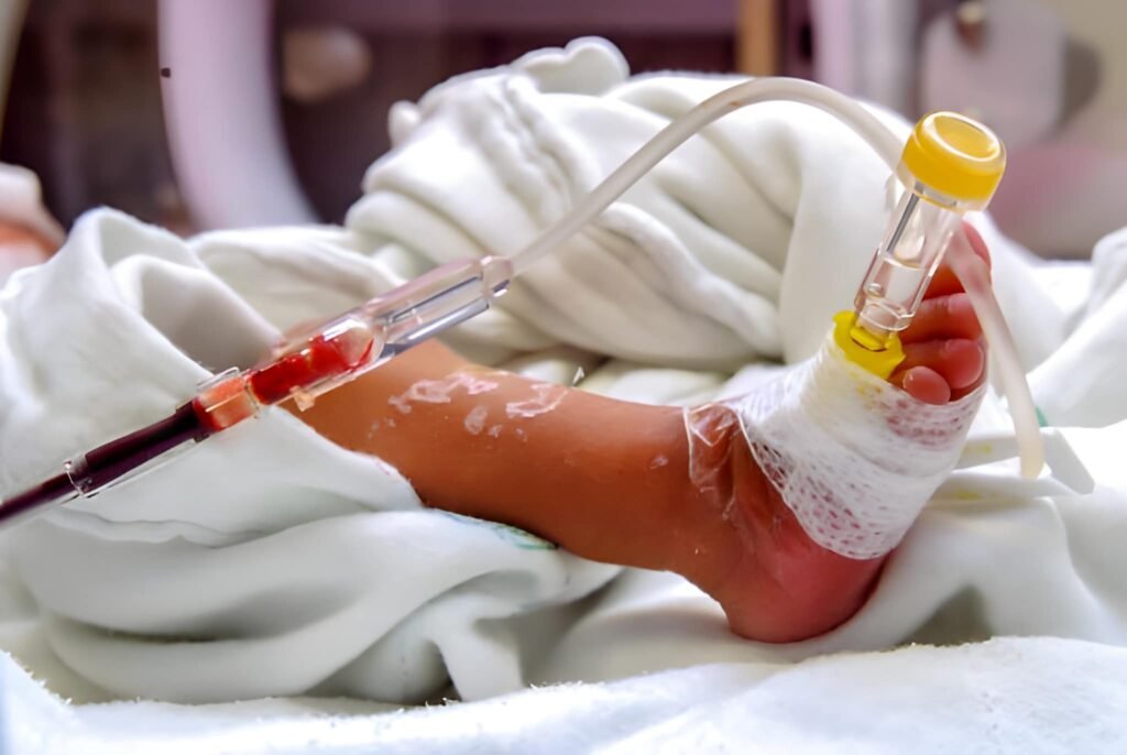
First, evaluate the baby's hemodynamic status. If the hematuria and melena have been significant, the baby may be anemic or in shock. Intravenous access is mandatory.
- Vitamin K Administration: 1 mg of Vitamin K1 should be given intravenously for active bleeding. While the IM route is standard for prophylaxis, the IV route acts faster in an emergency.
- Fresh Frozen Plasma (FFP): If the bleeding is life-threatening, FFP (10-15 mL/kg) provides an immediate source of active clotting factors while waiting for the Vitamin K to work in the liver.
- Packed Red Blood Cells: If the hematocrit has dropped significantly (e.g., below 30% in a symptomatic neonate), a blood transfusion may be required.
Calculations for Management
Consider a 3.5 kg newborn with active melena and an estimated 10% blood volume loss.
Total Blood Volume: 3.5 kg x 80 mL/kg = 280 mL
Estimated Loss: 28 mL
FFP Dose: 3.5 kg x 10 mL/kg = 35 mL
This 35 mL of FFP provides the necessary clotting factors to bridge the gap until the Vitamin K takes effect (usually 6 to 12 hours).
Comparison: VKDB vs. DIC
The management for these two conditions differs significantly. Using the table below, we can see why clinical context is vital.
| Feature | Vitamin K Deficiency (VKDB) | Disseminated Intravascular Coagulation (DIC) |
|---|---|---|
| Appearance | Often healthy and vigorous | Usually very ill, pale, or lethargic |
| Platelets | Normal | Low (Thrombocytopenia) |
| PT / aPTT | Both Prolonged | Both Prolonged |
| Fibrinogen | Normal | Low |
| Trigger | Nutritional / Absorption | Sepsis, Asphyxia, Trauma |
| Main Treatment | Vitamin K Replacement | Treat the underlying illness + Blood products |
Prevention and Long-term Perspectives
The occurrence of Classic VKDB in developed nations is rare, yet we have seen a slight uptick due to "refusal of prophylaxis." It is vital for specialists to educate parents on the necessity of the Vitamin K shot. Oral Vitamin K is available in some regions but is less reliable than the intramuscular injection, especially for preventing Late VKDB.
Monitoring for Complications
Once the initial bleeding (umbilicus, melena, hematuria) is controlled, the primary concern shifts to intracranial hemorrhage (ICH). Even if the baby looks fine, a head ultrasound or CT scan is often warranted if systemic bleeding was severe. ICH can lead to permanent developmental delays, seizures, or cerebral palsy if not identified.
Key Takeaways for Parents and Providers
- Early detection is key: Any blood in the stool or oozing from the cord after the first 24 hours requires an immediate ER visit.
- The "Shot" is standard: A 1 mg dose of Vitamin K1 at birth reduces the risk of bleeding by over 80 times compared to no treatment.
- Breastfeeding support: While breast milk is the gold standard for nutrition, its low Vitamin K content makes the birth injection non-negotiable for breastfed infants.


