Umbilical Cord Anatomy: The Significance of the Three-Vessel Structure
Understanding the Normal Blueprint and the Implications of a Two-Vessel Cord (SUA)
Table of Contents
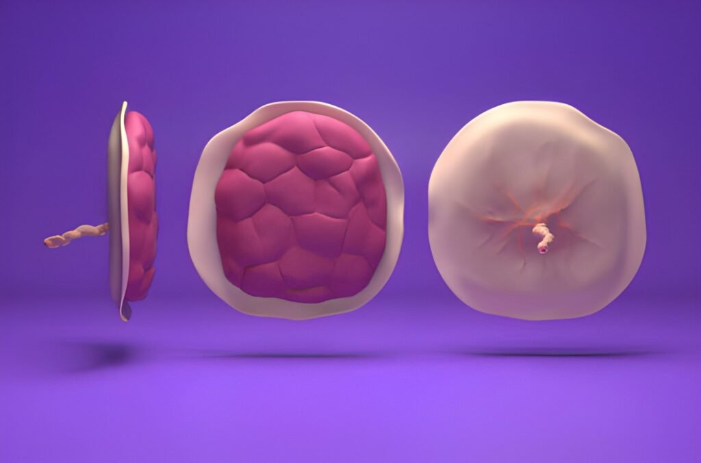
The umbilical cord is the lifeline connecting the developing fetus to the placenta, facilitating all gas exchange and nutrient transfer. When discussing the "three-vessel cord," it is crucial to recognize that this is the biological standard—the definition of a structurally normal umbilical cord. The true concern in prenatal diagnostics is usually the finding of a **two-vessel cord**, also known as a Single Umbilical Artery (SUA). This comprehensive guide clarifies the normal vascular blueprint, explains the implications of the anomaly, and details the rigorous monitoring steps that follow a diagnosis.
Normal Anatomy: The Three-Vessel Standard
A healthy umbilical cord contains three distinct blood vessels protected and encased within a gelatinous substance called Wharton’s jelly. These three vessels perform complementary, life-sustaining functions:
The Three Vessels of Life
- Two Umbilical Arteries: These carry deoxygenated blood and metabolic waste away from the fetus to the placenta. These vessels manage the waste disposal system.
- One Umbilical Vein: This carries oxygenated, nutrient-rich blood from the placenta directly to the fetus. This vessel manages the nourishment and breathing system.
This trio ensures a continuous, efficient, and balanced circulatory loop, allowing the fetus to thrive in the intrauterine environment.
The Anomaly: Single Umbilical Artery (SUA)
A Single Umbilical Artery (SUA), or two-vessel cord, is the most common structural variation of the umbilical cord. It is characterized by the absence of one of the two expected umbilical arteries, resulting in a cord containing only one artery and one vein. This finding occurs in approximately 0.5% to 6% of singleton pregnancies, making it a relatively frequent diagnosis on routine ultrasound.
How a Two-Vessel Cord is Diagnosed
The definitive diagnosis of a two-vessel cord is typically made during the Level 2 fetal anatomy scan, which is generally performed between 18 and 22 weeks gestation. During this scan, the technician uses color Doppler ultrasound to visualize the cross-section of the umbilical cord. A normal cord displays three distinct circles (the two arteries and one vein); an SUA cord displays only two.
Clinical Significance of a Two-Vessel Cord (SUA)
The diagnosis of an SUA can be concerning for parents, but the clinical significance depends heavily on whether it is an **isolated finding** or if it is associated with other structural anomalies.
Isolated Finding (Most Common Outcome)
In up to 75% of cases, the SUA is an isolated finding, meaning the fetus shows no other structural defects or genetic abnormalities. In these pregnancies, the remaining single artery typically enlarges to handle the full load of waste removal, and the pregnancy usually proceeds normally. The primary risk associated with isolated SUA is a slightly higher chance of fetal growth restriction, as the circulatory system is under increased pressure.
Association with Other Anomalies
When the SUA is not isolated, it can be linked to other structural issues, suggesting a broader developmental pathway anomaly. The systems most often implicated are:
- Renal (Kidneys and Urinary Tract): Kidney malformations or absent kidneys. The fetal circulation that forms the umbilical arteries is closely related embryologically to the structures that form the renal system.
- Cardiac (Heart): Structural heart defects. Since the umbilical vessels are central to fetal circulation, their malformation can sometimes coincide with abnormal heart development.
- Chromosomal: While rare, SUA is statistically associated with a higher risk of chromosomal abnormalities (e.g., Trisomy 18 or 13). However, if early genetic screening (NIPT) was negative, this risk is significantly mitigated.
Diagnostic Protocol and Monitoring
A diagnosis of SUA mandates a structured, higher level of prenatal surveillance to confirm whether the finding is isolated and to monitor the fetus’s growth and well-being.
Level 3 Ultrasound (Fetal Echocardiogram)
Upon an SUA diagnosis, the care provider will typically recommend a detailed Level 3 ultrasound, often including a dedicated fetal echocardiogram. This specialized scan provides an in-depth, high-resolution view of the fetal heart structure and function, thoroughly assessing for any associated cardiac defects.
Serial Fetal Growth Surveillance
The primary concern, even with an isolated SUA, is the risk of Intrauterine Growth Restriction (IUGR). To manage this risk, a revised monitoring schedule is established:
- Repeat Growth Scans: Quarterly or monthly ultrasounds are scheduled, beginning around 28 weeks, to measure fetal growth and ensure the single artery is efficiently supporting the pregnancy.
- Doppler Studies: These ultrasounds often include umbilical and fetal vessel Doppler studies to assess blood flow resistance and velocity, confirming optimal placental function.
This stringent monitoring allows the care team to intervene immediately if growth restriction is detected, ensuring the best possible outcome.
Interactive Risk Assessment Summary
Use this interactive guide to understand the risk summary based on whether the SUA is isolated or associated with other factors, allowing for confident discussion with your medical team.
Select the Status of the Two-Vessel Cord Finding:
Summary: Clinical Confidence
A "three-vessel cord" is the desired and normal anatomy, confirming robust circulation. If your prenatal care has identified the variant—a two-vessel cord (SUA)—it is essential to remain calm and follow the specialized monitoring plan. Since the majority of these findings are isolated, leading to a healthy outcome with appropriate surveillance, the focus shifts to ensuring optimal fetal growth through regular, specialized ultrasounds. Engage confidently with your Maternal-Fetal Medicine specialists to manage this condition and ensure comprehensive prenatal care.


